Αναζήτηση
ΚΛΕΙΣΙΜΟΦωτονική τεχνολογία για βιοϊατρικές εφαρμογές
Δρ. Χρήστος Ριζιώτης
Combining group’s expertise with practical needs from medical diagnostic and biomedical area has led also to a number of ongoing collaborations in applications like pancreatic islet/cell transplantation, antibiotic delivery, lab-on-chip applications, diagnostic microscopy etc.
In the last example it has been implemented the design and engineering of novel structures with high anticipated use in a number of microscopy based diagnostic techniques such as cytological PAP-Test. Cervical screening based on Papanicolaou (PAP) Test has been proved to be an efficient practice for preventing cervico-vaginal cancer in High Risk Human Papilloma Virus infected women. Introduction of liquid based process combined with automatization and digital image analysis has led to a more reliable diagnosis compared to conventional management of the slides. However, microscopy based diagnosis in Cytology is characterized by a variety of parameters, including specimens’ adequacy, inhomogeneous/discontinuous cell density, fixing/staining procedure quality, and also screeners’ experience and observation ability. Our group introduced a novel real-time screening aid and platform improving diagnosis in Pap -Test slides.
The proposed solution (patented) provides a customizable spatial grid with sequentially ordered and indexed segments than can be attached in different possible ways to the carrier platform (e.g. microscope slide) and can act as a calibration and orientation aid during the screening process. Such grids were developed by a number of technical means and also by Femtosecond Laser Micromachining technique on commercially available borosilicate glass based cover slips.
A set of difficult-borderline diagnostic PAP-Test cases were comparatively analyzed by conventional and grid based screening procedures, respectively. Statistical analysis showed that grid based microscopy led to a dramatically updated diagnosis by identifying a significantly increased number of abnormal cells in a shorter (4 instead of 5 min) screening time per slide compared to conventional microscopy, especially concerning the number of detected neoplastic/cancerous cells.
The grid can be fabricated and integrated in different platforms such as in solid glass cover slips, flexible polymers stripes and thin shelf-adhesive transparent tapes that could be attached afterwards on top of the slides. The grid has been applied, in collaboration with a number of diagnostic labs and university hospitals, both in pathology and also in cytology diagnostic cases such as ASCUS/ASC-H/LGSIL/HGSIL/AGC/INSITU ADENOCA/ADENOCA as well as in “In Situ Hybridization”- ISH techniques, and Immunocytochemistry-Immunohistochemistry (ICC-IHC) protocols for assessment of immunostained cell characteristics.
The capabilities and the high customization degree of the femtosecond laser micromachining system together with the adaptation of laser processing in various materials and photonic platforms have led to other advancements in biomedical technology.
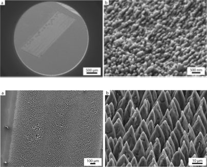
Femtosecond direct laser induced surface structuring on Silicon has led to customized surface’s nano/microtopography towards selective cancerous cells adhesion Our findings demonstrate that surfaces with microtopography are repellent, while those with nanotopography are attractive for MDA-MB-231 cell adherence leading thus to ways for the enhancement of diagnostic capability in relevant biomedical applications.
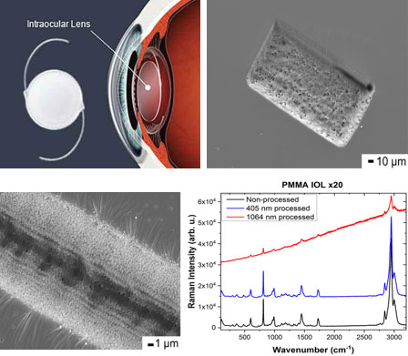
Polymeric Intraocular lenses (IOLs) are vital for restoring vision following cataract surgery and for correcting refractive errors. We employ laser micromachining technology for precise modifications via ablation on PMMA and acrylic IOLs, using femtosecond (fs), nanosecond (ns), and diode continuous wave (CW) lasers, at wavelengths ranging from near-ultraviolet to infrared. The results suggested the feasibility of accurate IOL patterning, which could offer personalized vision correction solutions, based on relevant corneal wavefront data, thus surpassing standard lenses, marking a significant advancement in cataract surgery outcomes.
Laser Processing of Intraocular Lenses
Applied Sciences, 2024, 14(14), 6071
Reference and Callibration Grid for Improved Real Time Detection of Biological Entities in Microscopy Diagnostic Techniques
Hellenic Industrial Property Organization, Patent No# 1008931
Key Publications

Anticancer Research 2020, 40, 3759
C. Riziotis, E. Tsiambas, “Reference and Calibration Grid for Medical Diagnostic Microscopy”, WIPO International PCT Patent Application, Patent Pending.
PCT/GR2016/000032, WO2017/009673.
ΟΒΙ. Hellenic Industrial Property Organization, Patent No#:1008931.